【概 要】
・糸球体基底膜とは、糸球体毛細血管の内皮細胞と足細胞の間にある厚い基底膜で、糸球体のろ過のしくみ(ろ過バリア)の中心となる膜である。
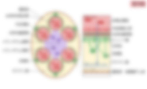
・毛細血管の内皮細胞の基底板と足細胞の基底板が融合してできたもので、ほかの組織の基底膜より厚い(約250~400nm)。基底膜の外側には、腎小体を包むボウマン嚢の臓側板をつくる足細胞が接している。
・電子顕微鏡では、内皮細胞側から順に、内透明層(lamina rara interna)・緻密層(lamina densa)・外透明層(lamina rara externa)の3層に見える。
・主にIV型コラーゲン、ラミニン、ヘパラン硫酸などを含む。内皮細胞と足細胞によってつくられ、保たれている。
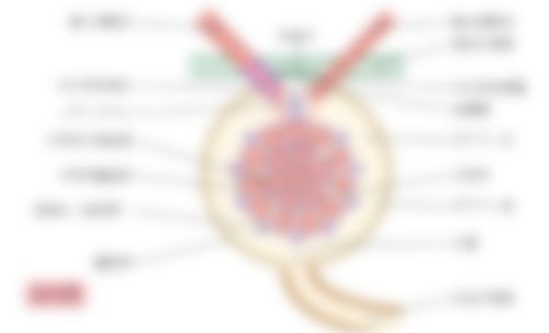
・原尿(ろ液)は、内皮細胞の小孔、この基底膜、足細胞の足突起の間のすき間(ろ過スリット)の順に通り、ボウマン腔へ出る。基底膜は大きさと電荷でふるい分けをし、アルブミンなどの血漿タンパク質を通しにくい。
・基底膜やろ過バリアが傷むと、タンパク質が尿にもれ出る(タンパク尿)。膜性腎症では基底膜が厚くなり、アルポート症候群ではIV型コラーゲンの遺伝子の異常で基底膜がもろくなる。
 この部位の閲覧には会員登録が必要です。
【参考となるサイト】
「 日本人体解剖学 」「 船戸和弥のHP 」
以下は「Wikipedia」の解説文となる。
The glomerular basement membrane of the kidney is the particular basement membrane of the glomerulus. The glomerular endothelial cells, the glomerular basement membrane, and the filtration slits between the podocytes perform the filtration function of the glomerulus, separating the blood in the capillaries from the filtrate that forms in Bowman's capsule. The glomerular basement membrane is a fusion of the endothelial cell and podocyte basal laminas, and is the main site of restriction of water flow. Glomerular basement membrane is secreted and maintained by podocyte cells.
【 語 句 】
・glomerular basement membrane:糸球体基底膜 ・basement membrane:基底膜 ・glomerulus:糸球体 ・endothelial cell:内皮細胞 ・filtration slit:ろ過スリット ・podocyte:足細胞 ・filtration function:ろ過の機能 ・capillary:毛細血管 ・filtrate:ろ液 ・Bowman's capsule:ボウマン嚢 ・fusion:融合 ・basal lamina:基底板 ・restriction:制限 ・secrete:分泌する ・maintain:維持する
■ 写真やイラストを掲載しているサイト ■
・ イラストや写真を掲載しているサイト-Ⅰ
・ イラストや写真を掲載しているサイト-Ⅱ
・ イラストや写真を掲載しているサイト-Ⅲ
・ イラストや写真を掲載しているサイト-Ⅳ